MycoplasmasFig2AFig2B.jpg
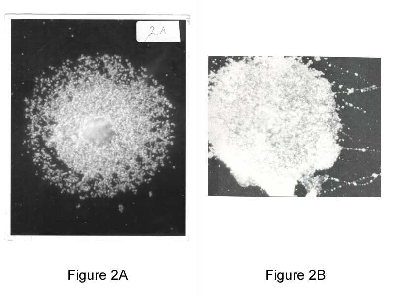
Image courtesy of The Arthritis Trust of America
Figure 2A (left): Mycoplasma Colony, Fixed on Glass Slide, 400X dark field. A dark-field photo of a mycoplasma colony showing the minimal reproductive particles.
Figure 2B (right): Dark-field photomicrographs of hot water-fixed Mycoplasma hominis, type 1, colonies, X 450. Variable large body and particulate chainlike filaments disrupted from colony. Shows the adhesiveness and pliability of mycoplasmas from a colony streaked on a slide producing filament like structures.