EntamoebaGingivalisMicroscopyColor.jpg
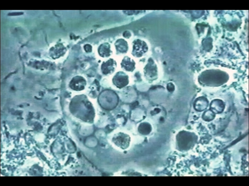
Image by Mark Bonner, DMD, Institut international parodontie; Wikimedia Commons / CC BY-SA 3.0
Entamoeba gingivalis from aggressive periodontal disease patient biofilm using phase contrast microscope 1000x. It is recognizable through its dense core in the middle, formed by a central point encircled by a circular halo and surrounded by bigger phagosomes inside a greyish cytoplasm.
