MycoplasmasFig5AFig5B.jpg

Image courtesy of The Arthritis Trust of America
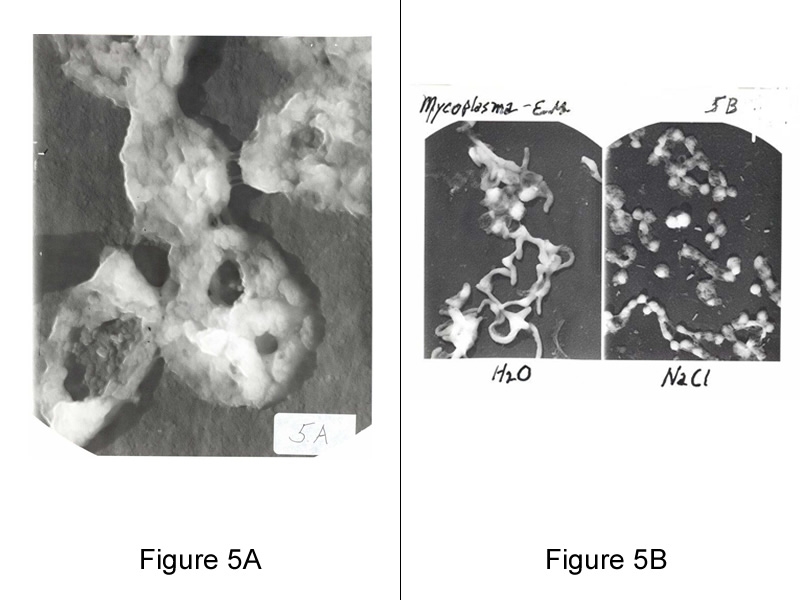
Figure 5A (left): Five Mycoplasmas: 120,000 X magnification by electronmicroscope. High magnification showing membrane surface with adhesive attachment and infrastructure.
Figure 5B (right): Mycoplasma in water and salt. Mycoplasma’s sensitivity to low osmolarity are morphology and viability dependent, showing coccoidal in isotonic saline, and filamentous in water.

